


HIDA Scan
A HIDA scan evaluates bile flow and gallbladder function when gallbladder symptoms persist or ultrasound is unclear. Learn how Dr. Colin Dunn uses HIDA results in Los Gatos for gallbladder evaluations.
What is a HIDA scan?
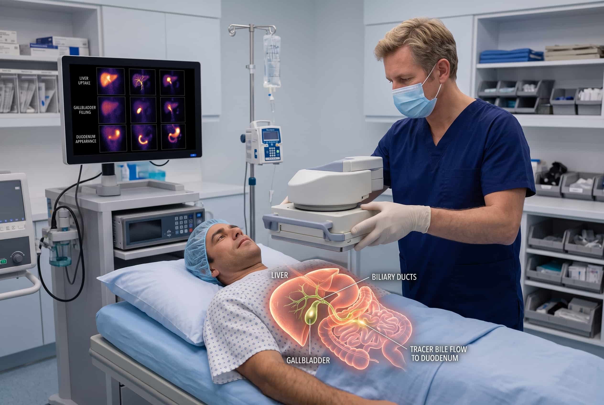
A HIDA scan is a nuclear medicine imaging test that evaluates how bile moves from the liver through the bile ducts, gallbladder, and small intestine. It is also known as a hepatobiliary scan or cholescintigraphy. This study tracks bile flow to show how well the biliary system is functioning. Unlike an ultrasound that shows anatomy, a HIDA scan focuses on how the gallbladder and bile system are working.

How a HIDA scan works
The test follows a small amount of radioactive tracer as it moves through the bile pathway from the liver to the small intestine.
What it measures
• Bile production and flow from the liver
• How well the gallbladder fills with bile
• The rate at which the gallbladder releases bile (ejection fraction)
• Movement of bile through the bile ducts and into the small intestine
What it tells Dr. Dunn
• Whether the gallbladder is functioning normally or emptying too slowly
• If there is a potential blockage in the cystic duct or bile ducts
• Whether symptoms like right upper abdominal pain are related to gallbladder function
• If a bile leak is present after a recent surgery or injury
Common uses for a HIDA scan
A HIDA scan provides functional information when symptoms, exams, or ultrasound results raise questions about the gallbladder or bile flow.
The scan measures the gallbladder ejection fraction to see if the gallbladder is emptying normally. This helps evaluate symptoms like pain after eating without visible stones, though a low ejection fraction does not automatically mean surgery is the right choice.
It can help identify acute or chronic inflammation of the gallbladder, especially when an ultrasound is not definitive or when the cystic duct might be blocked.
The test can detect if bile is leaking outside the normal biliary system after a surgical procedure or an injury.
What to expect during your HIDA scan
The scan is performed by a radiology or nuclear medicine team and involves several steps to track the movement of the tracer.
During diagnostic endoscopy
• A small amount of radioactive tracer is injected through an IV into your bloodstream.
• A gamma camera takes images as the tracer moves from your liver into the gallbladder and intestines.
• You may receive a medication called sincalide to make your gallbladder contract and empty for measurement.
• The imaging process can take one to four hours depending on how quickly the tracer moves.
During surgery
A HIDA scan is not performed during surgery. However, the results are used before surgery to help Dr. Dunn plan a laparoscopic cholecystectomy or other gallbladder procedures. In some cases, a HIDA scan may be used after surgery to check for bile leaks if a patient has new or worsening symptoms.
Patients often seek a HIDA scan when they experience persistent right upper abdominal pain, nausea after meals, or bloating that ultrasound does not fully explain.
Why a HIDA scan matters for your care
The value of a HIDA scan comes from interpreting the functional results in the context of your overall health. Dr. Dunn reviews these findings alongside your symptoms, physical exams, ultrasound results, and blood tests. This comprehensive approach helps determine if your symptoms are truly related to your gallbladder and if surgery is the most appropriate next step for your recovery.
Want to understand how this applies to your case?
Book a consultation and Dr. Dunn will walk you through exactly how EndoFLIP is used in your specific planned procedure.
Procedures informed by HIDA scan results
HIDA scan results help guide the decision-making process for various gallbladder and biliary treatments.
Used in these procedures
• Laparoscopic cholecystectomy (gallbladder removal)
• Evaluation of biliary dyskinesia
• Assessment of acute or chronic cholecystitis
• Post-surgical bile leak evaluation
Not used in these procedures
A HIDA scan is not typically the first imaging test for gallbladder pain and does not replace a gallbladder ultrasound. It is also not used during pregnancy, and breastfeeding patients may need to discard pumped milk for 24 hours after the scan. Certain opioid pain medications may also be withheld because they can affect bile flow.
Frequently Asked Questions

A HIDA scan is a nuclear medicine imaging test used to evaluate the function of the liver, gallbladder, and bile ducts. A small amount of radioactive tracer is injected into a vein and tracked by a special camera as it moves through the biliary system. This study is often recommended when symptoms continue but initial tests like ultrasound are unclear or normal.
You will typically be asked to fast for several hours before the appointment because eating can change how the gallbladder functions during the test. It is important to review all current medications and supplements with the care team as some can affect bile flow. Patients should also inform the imaging staff if they are pregnant or breastfeeding before the procedure begins.
After a radioactive tracer is injected into a vein, a gamma camera captures images of the abdomen to record the movement of bile over time. In some cases, a medication is given to stimulate the gallbladder to contract so the imaging team can measure how much bile is released. Some patients may experience temporary upper abdominal cramping if this stimulating medication is used.
The scan shows whether bile is moving normally through the biliary pathway and how well the gallbladder fills and empties. Findings such as a low ejection fraction or delayed tracer movement may suggest gallbladder dysfunction or chronic inflammation. Dr. Dunn reviews these functional results alongside your symptoms, physical exam, and other imaging to determine the most appropriate next step.

