
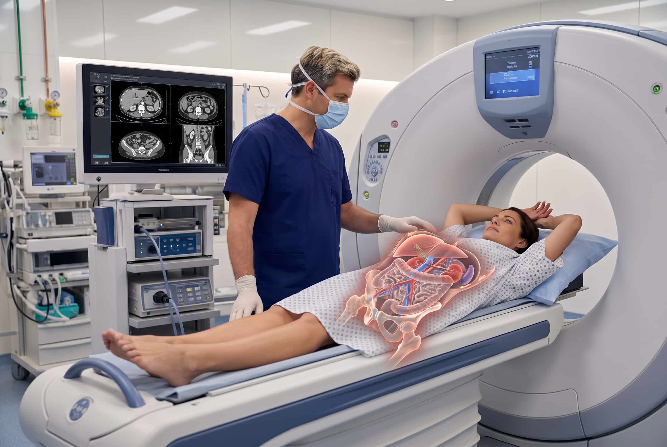


CT Abdomen & Pelvis
Learn how a CT abdomen and pelvis scan helps evaluate abdominal pain, diverticulitis, and hernias to support surgical planning with Dr. Colin Dunn.
What is a CT abdomen and pelvis scan?
A CT abdomen and pelvis scan is a diagnostic test that uses X-rays and computer processing to create detailed pictures of the belly and pelvic area. These images show internal organs, bones, soft tissue, and blood vessels in greater detail than a standard X-ray. The scan takes many image slices that can be viewed from different angles to help your doctor see what is happening inside your body.

How a CT scan works
The CT scanner uses X-rays and computers to take many detailed pictures of your body in slices. This allows the doctor to see your organs and tissues from different angles, which is more detailed than a standard X-ray.
What it measures
• Internal organs like the liver, kidneys, and pancreas
• The small and large intestines (bowel)
• Blood vessels and lymph nodes
• Abdominal wall and hernia locations
• Signs of inflammation, infection, or fluid collections
• Masses, tumors, or kidney stones
What it tells Dr. Dunn
• If there is a structural cause or signs of inflammation
• The exact location and severity of a condition like diverticulitis
• Whether surgery, medication, or more testing is the best next step
• How complex a surgical repair might be based on your body's structure
• If an urgent evaluation or hospital care is needed
Common uses for CT imaging
A CT scan may be recommended to evaluate abdominal pain, infections, hernias, or to help plan for a surgical procedure.
CT scans help find the cause of pain in different areas of the belly by looking at multiple organs and structures in one exam.
Imaging can confirm if the colon is inflamed, locate the affected area, and check for complications like an abscess.
A scan can show the size of a hernia, what is inside it, and help the surgeon plan the best way to repair it.
What to expect during your scan
A CT scan is usually a fast and painless process where you lie on a table that slides into the scanner.
During diagnostic endoscopy
• You will receive instructions on whether to fast or if contrast is needed.
• You may be given contrast material by mouth or through an IV to highlight certain areas.
• Patients with kidney disease or diabetes may need blood tests before receiving IV contrast.
• You will lie on a narrow table, usually on your back, and slide into the scanner.
• You may be asked to hold your breath briefly while the images are taken.
• The entire scanning process usually takes less than 30 minutes.
• You should immediately report any trouble breathing, hives, or severe itching during or after the scan.
During surgery
A CT scan is not typically performed during a surgical procedure. Instead, Dr. Dunn uses the images from the scan before surgery to understand your specific anatomy and plan the most effective approach for your operation.
Patients often want to know what their imaging results mean in plain language. Dr. Dunn reviews your scan with you to explain the findings and discuss whether surgery is the safest and best next step for your health.
Why this imaging matters
A CT scan provides important information, but it is just one part of your evaluation. Dr. Dunn reviews these images along with your symptoms, physical exam, and medical history to create a safe and personalized care plan. This helps determine if surgery is the best option for you.
Want to understand how this applies to your case?
Book a consultation and Dr. Dunn will walk you through exactly how EndoFLIP is used in your specific planned procedure.
Which conditions use this scan?
CT imaging is used to evaluate many conditions, including abdominal pain, diverticulitis, hernias, and cancer workup.
Used in these procedures
• Abdominal or pelvic pain
• Diverticulitis and its complications
• Appendicitis
• Abdominal wall or groin hernias
• Bowel obstructions or inflammation
• Cancer workup and staging
• Kidney stones
Not used in these procedures
A CT scan is not right for every symptom and does not replace other tests like an ultrasound or endoscopy. For example, an ultrasound is often better for certain gallbladder issues, and a colonoscopy is needed to see the inner lining of the colon. Because CT scans use a small amount of radiation, Dr. Dunn will determine if a CT scan is the most appropriate and safest next step for your specific situation.
Frequently Asked Questions

A CT abdomen and pelvis scan is a diagnostic imaging test that uses X-rays and computer processing to create detailed cross-sectional images of the belly and pelvic area. These images provide greater clarity than traditional X-rays for evaluating organs, bones, soft tissues, and blood vessels. Dr. Dunn uses these results to help identify causes of abdominal pain, infection, or inflammation and to plan surgical care when necessary.
Preparation depends on whether your scan requires contrast material to highlight specific internal structures. You should wear comfortable, loose clothing and may be asked to fast for a few hours before the exam if IV contrast is used. Patients with certain conditions, such as kidney disease or diabetes, may need recent blood tests to ensure contrast can be used safely.
A CT scan is usually a fast and painless experience where you lie on a table that slides into the center of the scanner. The scanning portion of the appointment typically takes less than 30 minutes, though you may be asked to hold your breath briefly for clear images. If IV contrast is used, you might notice a brief warm flushing sensation or a metallic taste during the procedure.
Dr. Dunn reviews the radiologist's report and the images alongside your physical exam findings and medical history. This comprehensive review helps determine whether your condition requires surgery, further testing, or nonoperative management. Once the review is complete, you will meet to discuss the findings and establish a personalized treatment plan.

